
www.industry-asia-pacific.com
15
'26
Written on Modified on
ZEISS Unveils VersaXRM 5 Insight for Hybrid 3D X-Ray Imaging
Hybrid platform combines X-ray microscopy and microCT with AI-enabled reconstruction, supporting multi-scale, non-destructive imaging for research, inspection and life sciences.
www.zeiss.com
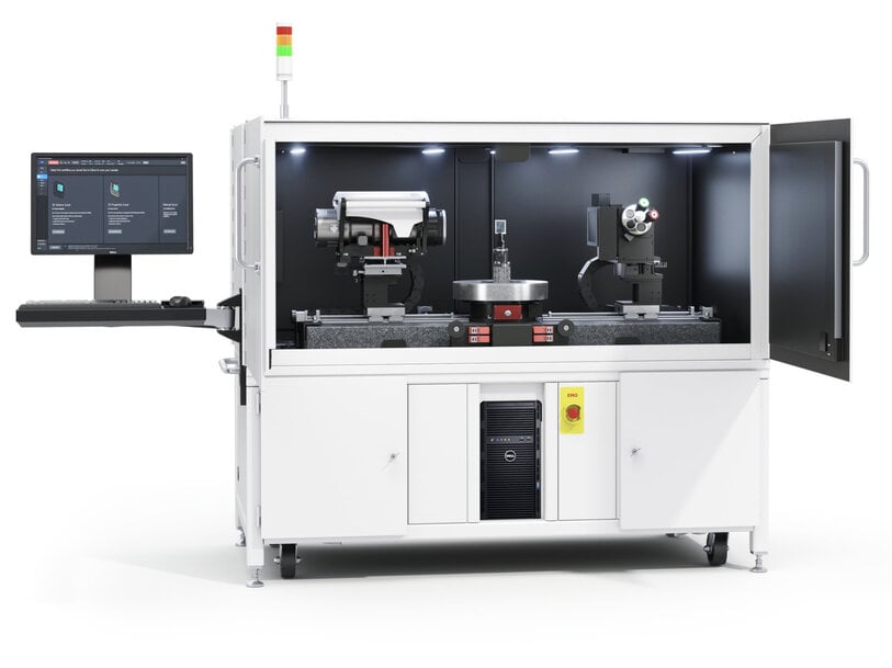
ZEISS has introduced the VersaXRM 5 Insight, a hybrid three-dimensional X-ray imaging platform combining computed tomography throughput with sub-micron microscopic resolution. The system targets structural engineering, electronic package inspection, additive manufacturing, and life sciences research by enabling multi-scale volumetric characterization within an uninterrupted, single-instrument workflow.
Multi-Scale Non-Destructive Volumetric Characterization
Engineering and scientific evaluation of structural components, composite materials, and biological tissues typically requires assessing both broad macrostructures and localized sub-micron defects. Conventional non-destructive evaluation workflows often compel operators to choose between wide-field micro-computed tomography (microCT) units and separate high-magnification X-ray microscopes (XRM). Alternating between discrete systems requires specimen extraction, re-fixturing, and manual spatial co-registration, introducing preparation errors and operational delays.
The VersaXRM 5 Insight consolidates wide field-of-view scanning and localized high-resolution imaging into a single instrument architecture. Operators can perform macroscopic screening across an entire component to detect structural anomalies, define targeted volumes of interest, and conduct deep volumetric scans without altering physical hardware setups or modifying test environmental chambers.
Preserving Specimen Integrity Through Resolution at a Distance
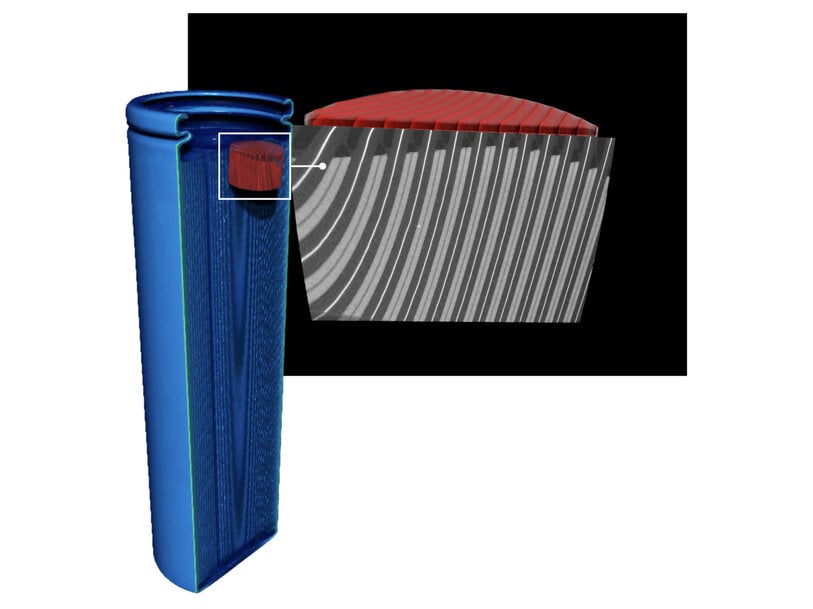
A major physical constraint in laboratory X-ray computed tomography is geometric magnification, where high resolution typically requires positioning the sample in extreme proximity to the X-ray source. This requirement often necessitates mechanical trimming, destructive cross-sectioning, or physical coring of large workpieces to prevent physical collisions with the source or detector.
The system incorporates Resolution at a Distance (RaaD) architecture, which utilizes dual-stage optical magnification downstream of the scintillator. This enables sub-micron spatial resolution while maintaining large working distances. Engineers can inspect critical defects, void distributions, and weld porosity inside bulky parts, intact electronic housings, and in situ mechanical test cells without mechanical modification, preserving sample structure for longitudinal four-dimensional testing.
Automated Navigation and Accelerated Acquisition Workflows
Operator variability, system alignment, and volume reconstruction times represent common bottlenecks in volumetric X-ray workflows. The VersaXRM 5 Insight incorporates ZEN navx control software to deliver automated, sample-aware navigation and procedural guidance, preventing collision trajectories between specimen fixtures and instrument components.
To accelerate experimental iteration, the platform integrates FAST Mode, which delivers rapid feedback for spatial alignment, macroscopic component screening, and intermediate-resolution tomographic reconstructions. The system also embeds artificial-intelligence algorithms within the reconstruction pipeline to mitigate beam hardening, reduce ring artifacts, and accelerate volume rendering from fewer angular projections.
Dr. Keith Duncan, Research Scientist and Director of X-ray Imaging at the Danforth Plant Science Center, noted that the detector architecture combines a wide-area flat-panel detector with secondary optical magnification to transition from global specimen survey to localized structural detail without mechanical reconfiguration.
Nicolas Gueninchault, Head of the X-ray Microscopy Field of Business at ZEISS Microscopy, indicated that integrating high-throughput microCT hardware with X-ray microscope optics balances spatial resolution, data acquisition speed, and operational usability on a single chassis.
Targeted Industrial and Materials Research Applications
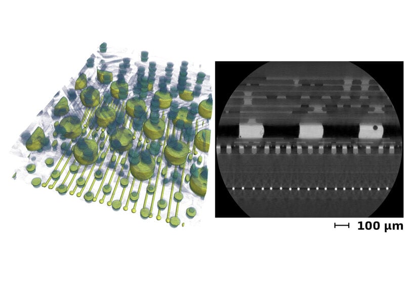
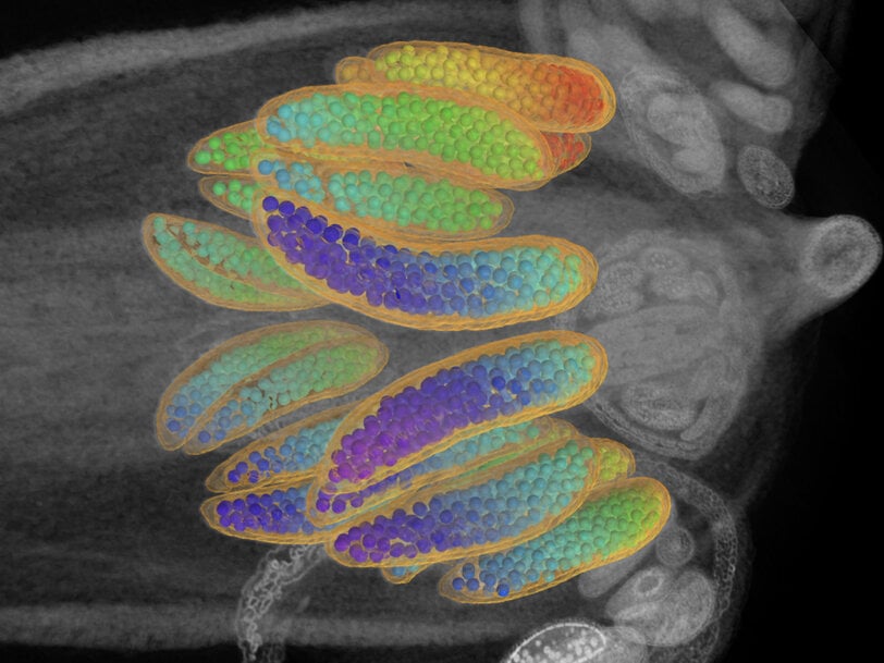
In materials science, the system enables non-destructive visualization of crack propagation, micro-void coalescing, and particulate dispersion during environmental and mechanical loading stages. In industrial microelectronics and semiconductor manufacturing, the instrument supports non-destructive root-cause analysis of solder ball fatigue, silicon via bridging, wire bond detachment, and internal delamination within packaged integrated circuits. Life sciences facilities deploy the platform for non-destructive, three-dimensional histological modeling and morphological phenotyping of intact plant and animal specimens.
Additional Context:
This section details technical specifications and competitive benchmarking not included in the original product announcement
The hybrid architecture of the ZEISS Versa series pairs a sealed microfocus X-ray source (typically operating between 30 and 160 kilovolts with an output up to 10 watts) with a dual-stage magnification mechanism. The primary detection path employs a high-resolution flat-panel CMOS detector for macroscopic screening, supplemented by an automated objective turret housing optical objectives (such as 0.4x, 4x, 20x, and 40x magnification) coupled to high-efficiency scintillators and scientific CCD or CMOS cameras. This configuration achieves true spatial resolutions down to 500 nanometers and minimum voxel dimensions down to 40 nanometers while supporting source-to-sample distances exceeding 20 to 50 millimeters.
Direct industry benchmarks for multi-scale non-destructive 3D X-ray systems include the Bruker SkyScan 2214 and the TESCAN UniTOM XL.
The Bruker SkyScan 2214 utilizes an open-type nanofocus X-ray source with diamond windows capable of beam energies up to 160 kilovolts, generating spot sizes below 500 nanometers. It incorporates modular detection options, combining an 11-megapixel cooled CCD camera for sub-micron evaluation with a 6-megapixel flat-panel detector for large-object overview (up to 300 millimeters in diameter). Unlike the optical magnification stages utilized in RaaD architecture, the SkyScan 2214 relies primarily on high geometric magnification, requiring the sample to sit within 1 to 2 millimeters of the source to achieve its maximum spatial resolution of 500 nanometers, which restricts high-magnification scanning of large or encased specimens.
The TESCAN UniTOM XL is engineered for high-throughput multi-scale microCT, integrating dynamic continuous scanning capabilities and supporting sample payloads up to 10 kilograms within a 300-millimeter field of view. The UniTOM XL operates a high-power microfocus X-ray source up to 160 kilovolts and 200 watts, utilizing a high-speed flat-panel detector that enables temporal resolution for dynamic 4D computed tomography down to sub-second volume acquisition. While delivering superior frame rates and volumetric throughput for larger engineering castings and dynamic mechanical compression cycles, its purely geometric magnification configuration limits maximum spatial resolution to approximately 3 to 4 micrometers, making it less specialized for sub-micron void and internal boundary isolation than multi-stage optical systems.
Edited by Natania Lyngdoh, Induportals editor, assisted by AI.
www.zeiss.com
